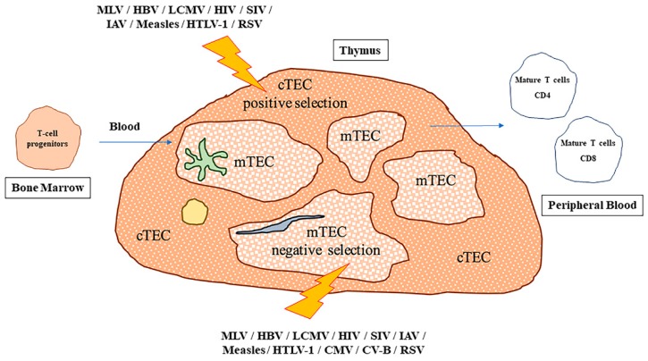
Figure 1.
Impact of viral infections on intra-thymic T cell development. Two major populations of thymic epithelial cells (TECs) are present in the thymic parenchyma. In particular, cortical TEC (cTEC) cells are identified in the cortical compartment of the parenchyma, whereas medullary TEC (mTEC) cells are located internally. Dendritic cells (DCs, shown in green), fibroblasts (blue) and macrophages (yellow), contribute to the tissue organization. In humans, during the maturation process, bone marrow-derived T-cell progenitors arrive in the thymus via the bloodstream, where cTEC cells contribute to positive selection maturation, and mTEC cells contribute to the negative selection process, respectively. Also, DCs are likely responsible for negative selection in the cortex. Once the selection has been successfully completed and self-reactive cells have been removed by apoptosis, mature CD4+ or CD8+ T cells leave the thymus to populate the peripheral blood and secondary lymphoid organs. Viral infections are able to affect thymus functionality in multiple ways, but the most severe effects are observed when positive or negative selection is impaired, as these processes drive key steps of T-cell development. In the figure, lightning bolts indicate which viruses can specifically target and influence the activity of cTEC or mTEC populations.

