Fig. 6.
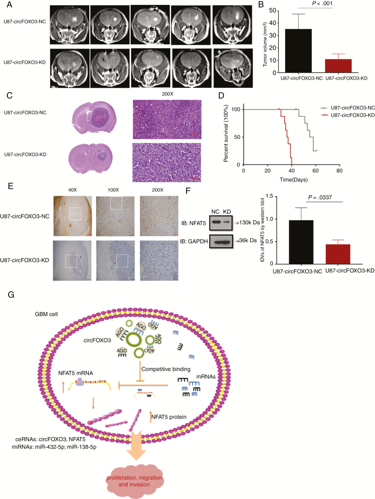
CircFOXO3 KD inhibits GBM progression in vivo. (A) Representative MR images of xenograft GBM tumors orthotopically inoculated with cells with NC or circFOXO3 KD on 3 weeks post-implantation. (B)Tumor volumes were calculated for each group (n = 8 per group). (C) Representative hematoxylin and eosin images of each group are shown (magnification: 200×; scale bar = 100 μm). (D) The comparative survival of mice bearing NC or circFOXO3 KD tumors was determined. The time of death was recorded as days after U87-MG implantation. (E) Representative immunohistochemistry images of NFAT5 in tumors collected from each group (magnification: 40×, 100×, 200×; scale bar = 500 μm, 200 μm, 100 μm). (F) Western blot analysis of NFAT5 in tumor collected from each group was performed. A multiple comparisons test adjusted P-value of <0.05 was considered statistically significant. The error bars represent the mean ± SD. (G) Model for the mechanism of circFOXO3 in GBM. CircFOXO3 sponges miR-138-5p/miR-432-5p through MREs and thus acts as a ceRNA to regulate NFAT5 expression and modulate tumor cell function in vitro and in vivo.

