Fig. 1.
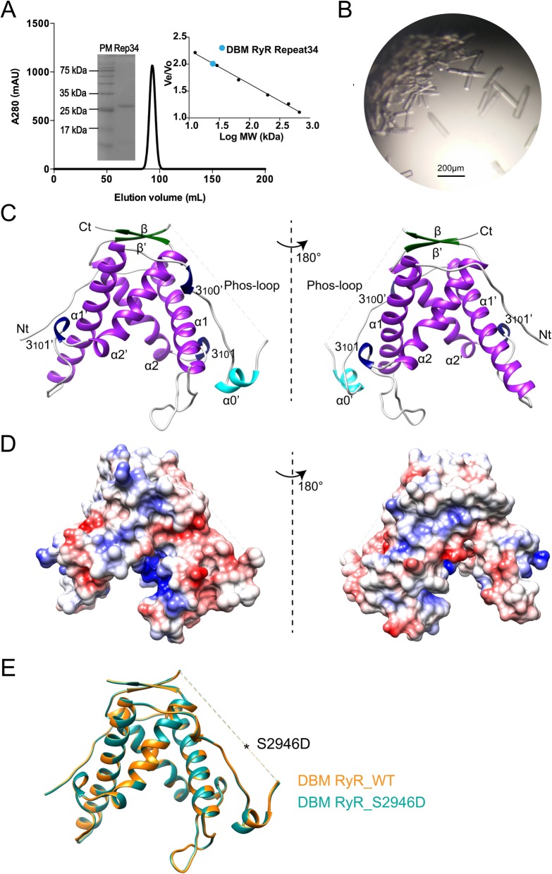
Structure of DBM Repeat34. a Elution profile of DBM RyR Repeat34 by gel-filtration chromatography using a Superdex 200 26/600 column (GE Healthcare, USA). The right inset shows the plotted standard curve for this column. The molecular weight estimated from its elution volume is ~ 28.1 kDa, suggesting monomeric form in solution (predicted MW is 24.9 kDa). The left inset is a 15% SDS-PAGE of purified DBM RyR Repeat34 showing protein marker (PM) in the left lane and purified Repeat34 (Rep34) in the right lane. b DBM RyR Repeat34 crystals produced by the hanging-drop method. c The crystal structure of DBM RyR Repeat34 in two different views are colored according to their secondary structure elements: α helixes in purple, 310 helixes in blue, β strands in green, and loops in gray. The Lepidoptera-specific helix α0′ is highlighted in cyan. d Electrostatic surface views of DBM RyR Repeat34. Negatively charged, positively charged, and non-charged surfaces are colored in red, blue, and white, respectively. e Structural superposition of the wild type and the S2946D mutant of DBM RyR Repeat34. The phospho-mimetic mutation is located in a flexible loop shown as dash line

