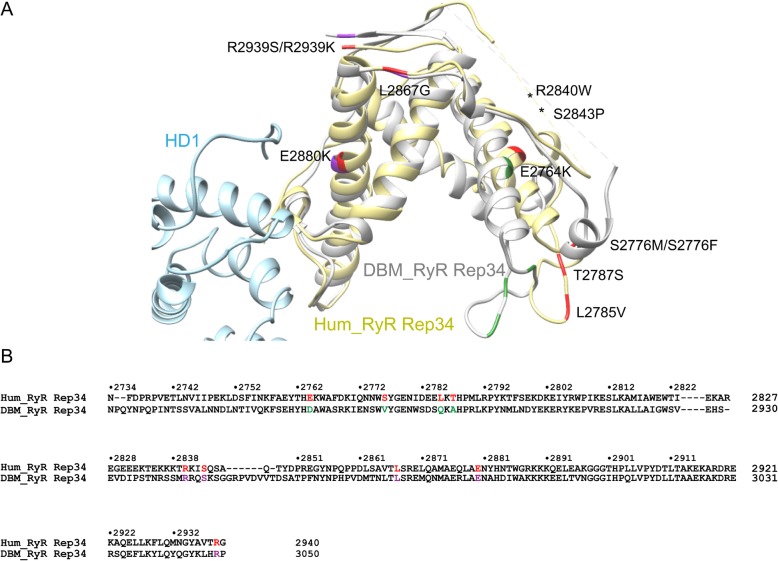
Fig. 8.
Disease-associated mutations mapped onto DBM Repeat34. a The crystal structure of DBM RyR Repeat34 is superposed with RyR1 cryo-EM model (PDB ID 5TAQ). Disease-associated mutations are colored in red in RyR1 structure. Corresponding conserved and non-conserved residues in DBM Repeat34 are colored in purple and green, respectively. b Sequence alignment between human RyR1 Repeat34 and DBM RyR Repeat34. The color scheme for disease mutations is the same as in panel A

