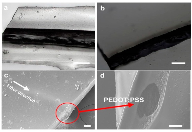
Figure 5.
(a,b) Optical imaging of upper and lower surfaces, showing the sheathed dark conducting PEDOT:PSS wire enclosed in the transparent fibroin sheath. (c) SEM imaging of the sheathed wires. (d) Cross-section across the wire showing the dark fiber surrounded by the insulating silk matrix. Scale bar = 50 µm in all panels.

