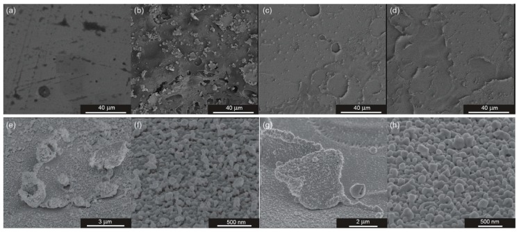
Figure 4.
Scanning electron microscopy (SEM) images of PET/ITO foil in few different states of SERS platform preparation: (a) raw PET/ITO foil without any modifications, (b) foil after 90 s of the DBD, (c) foil modified with DBD and with 30 nm layer of silver, (d) foil modified with DBD and with 70 nm layer of silver, (e,f) images of foil modified with DBD and with 30 nm layer of silver at high magnifications, and (g,h) images of foil modified with DBD and with 70 nm layer of silver at high magnifications.

