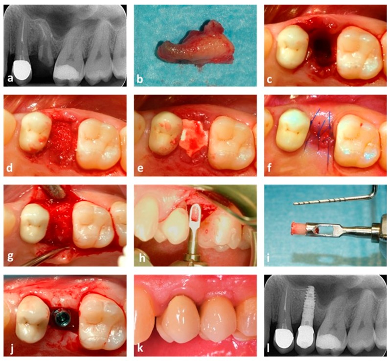
Figure 4.
Post-extractive socket grafting. A case where ABB was used is shown. (a) X-ray showing the initial patient status. Tooth 25 is fractured and compromised; (b) the element after being extracted; (c) the post-extractive socket before grafting; (d) the socket after being grafted; (e) a collagen membrane is placed below the gingival rims to cover the graft; (f) single cross stitches stabilize the reconstruction; (g) appearance of the regenerated socket 5.3 months after the grafting surgery; (h) a bone core is collected using a trephine bur; (i) the bone sample after collection; (j) an implant (Stone, 3.75 × 14 mm, IDI Evolution, Concorezzo, Italy) is placed into the regenerated bone; (k) the final prosthetic rehabilitation; (l) one-year control radiograph, showing maintenance of peri-implant bone levels.

