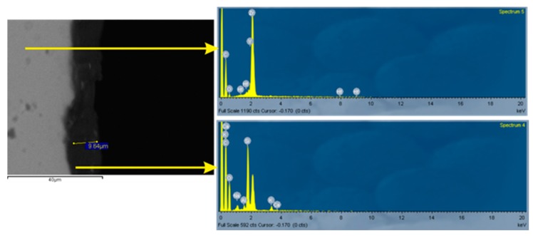
Figure 3.
SEM atomic-number contrast backscattered electron image of a cross-sectioned glazed monolithic zirconia specimen and associated EDS analyses. Dispersed phases with a grayscale level matching that of the substrate are apparent in the glaze film. Top EDS spectrum: ground zirconia substrate, bottom EDS spectrum: glaze layer.

