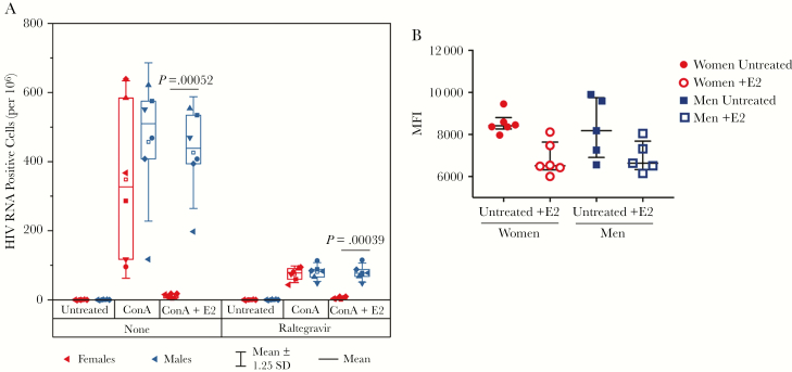
Figure 3.
Estrogen blocks human immunodeficiency virus (HIV) RNA transcription and spreading infection. A, Isolated CD4+ T cells from 6 male (blue symbols) and 6 female (red symbols) donors were stimulated with concanavalin A and cultured in the presence or absence of 1 μM raltegravir with and without 300 pg/mL 17β-estradiol. The number of HIV RNA–positive cells per million was quantified via envelope detection by induced transcription-based sequencing after 9 days of culture. Open symbols, median; horizontal line, mean; box plots, interquartile range; whiskers, ±1.25 standard deviations. Statistical comparisons were made with unpaired t test with Welch correction, and significant values are indicated on the graph. B, Peripheral blood mononuclear cells from men (n = 5) and women (n = 6) were cultured with and without 300 pg/mL 17β-estradiol (denoted as +E2 in the figure), and estrogen receptor 1 expression was measured in CD4+ T cells with intracellular staining and quantified by geometric mean fluorescence intensity. There was no statistically significant difference in untreated cells or after culture with 17β-estradiol (median and interquartile ranges are shown, Mann–Whitney statistics). Abbreviations: ConA, concanavalin A; HIV, human immunodeficiency virus; MFI, mean fluorescence intensity; SD, standard deviation.

