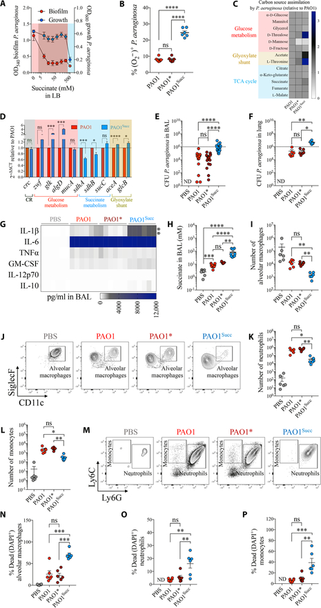
Fig. 3. Succinate promotes metabolic adaptation of P. aeruginosa and airway infection.
(A) Growth and biofilm of WT P. aeruginosa (PAO1) in increasing concentrations of succinate in LB media (n = 3). Pink, no succinate; red, 50 to 250 mM succinate; blue, 500 mM succinate. (B) Intracellular O2*−2 (ROS) in P. aeruginosa by succinate (PAO1, no succinate; PAO1*, 50 mM succinate; PAO1Succ, 500 mM succinate) (n = 3). (C) Relative single-carbon source assimilation by PAO1 (control), PAO1*, and PAO1Succ (n = 3). (D) mRNA expression relative to PAO1 for different metabolic pathways (n = 3). Mice were either infected with PAO1, PAO1*, or PAO1Succ or treated with phosphate-buffered saline (PBS). Twenty-four hours later, the following were analyzed in mouse airways: (E and F) CFU found in BAL and lungs (6 to 18 mice per group pooled from n = 3), (G) heatmap of different cytokines accumulated in BAL (6 mice per group pooled from n = 3), (H) succinate accumulated in BAL (5 to 11 mice per group pooled from n = 3), (I and J) number and density plots of viable alveolar macrophages in BAL (6 mice per group pooled from n = 3), (K to M) number and density plots of viable neutrophils and monocytes in BAL (6 mice per group pooled from n = 3), and (N to P) percentage of dead cells in BAL (6 mice per group pooled from n = 3). Data are shown as means ± SEM. ****P < 0.0001; ***P < 0.001; **P < 0.01; *P < 0.05; one-way ANOVA. ND, not detected. TNFα, tumor necrosis factor–α; GM-CSF, granulocyte-macrophage colony-stimulating factor; DAPI, 4′,6-diamidino-2-phenylindole.

