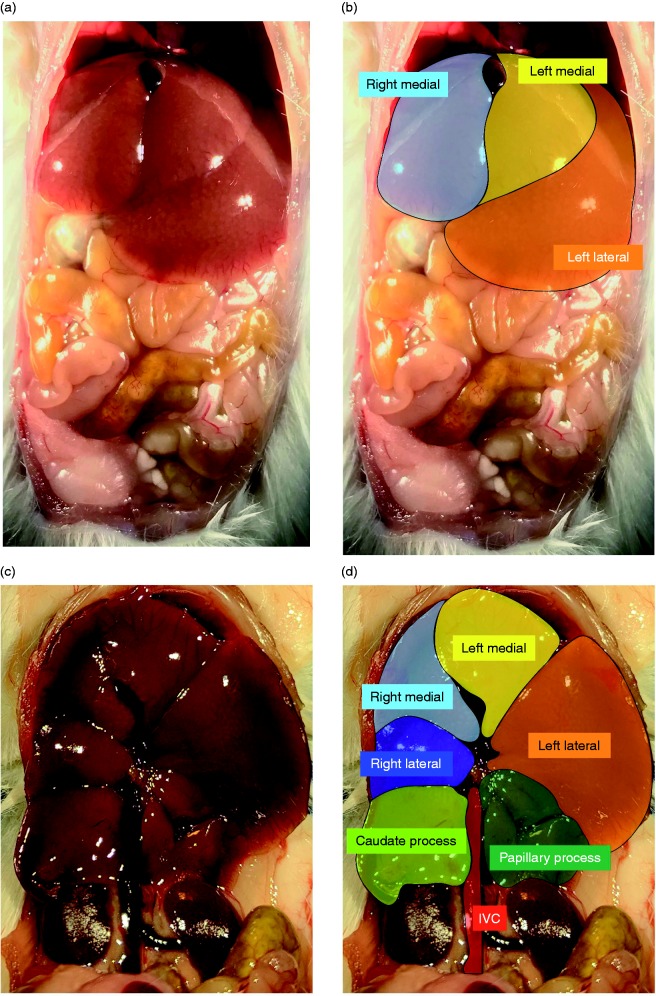
Figure 1.
Murine liver lobes as assessed in the current study, as adapted from Fiebig.22 (a) Ventral view of a male ICR mouse liver with corresponding color coding (b) showing the right medial lobe (light blue), left medial lobe (yellow) and left lateral lobe (orange). (c) Ventral view of the male ICR mouse liver with the stomach, small, and large intestine removed and corresponding color coding (d) right medial lobe (light blue), left medial lobe (yellow), right lateral lobe (dark blue), left lateral lobe (orange), caudate process (light green), and papillary process (dark green).

