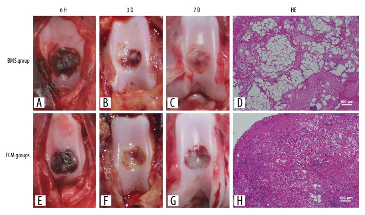
Figure 2.
Macroscopic appearance and histology of the area of exudate of the healing cartilage defect in the autologous bone mesenchymal stem cell (MSC)-derived extracellular matrix (ECM) scaffolds after bone marrow stimulation. The macroscopic appearance showed that the exudate of the healing wound in the bone marrow stimulation (BMS) group (A–C) did not organize well, whereas those in the extracellular matrix (ECM) group (E–G) are undergoing fibrosis. Histology shows that more chondrocytes were evenly distributed in the ECM group (H). The cells show good adhesion to the wall of the scaffold when compared with the BMS group (D).

