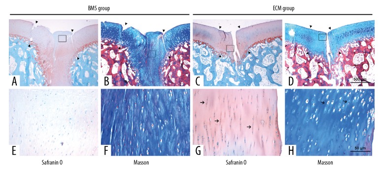
Figure 5.
Photomicrographs of the histology show the Safranin-O and Masson’s trichrome staining of the repaired cartilage of the bone marrow stimulation (BMS) group and the extracellular matrix (ECM) group at 6 months after surgery. Safranin-O staining showed that the repaired tissue in the bone marrow stimulation (BMS) group (A, E) showed poor metachromatic staining and was different from the normal surrounding cartilage and showed clear demarcation (arrowhead). However, the repaired tissue in the extracellular matrix (ECM) group (C, G) showed good integration with the surrounding cartilage (arrowhead), and the intensity of metachromatic staining resembled that of normal cartilage. The chondrocytes formed mature lacunae and were perpendicularly aligned (arrow). Masson’s trichrome staining showed that the defects were partially filled with fibrous tissue in the BMS group (B, F), with large numbers of rounded cells embedded in a fibrous and organized ECM. In contrast, the repaired tissue in the ECM group (D, H) showed several clusters of chondrocyte-like cells (arrow). Growth plates were observed in some zones. (A–D) Magnification ×10. (E–H) Magnification ×400.

