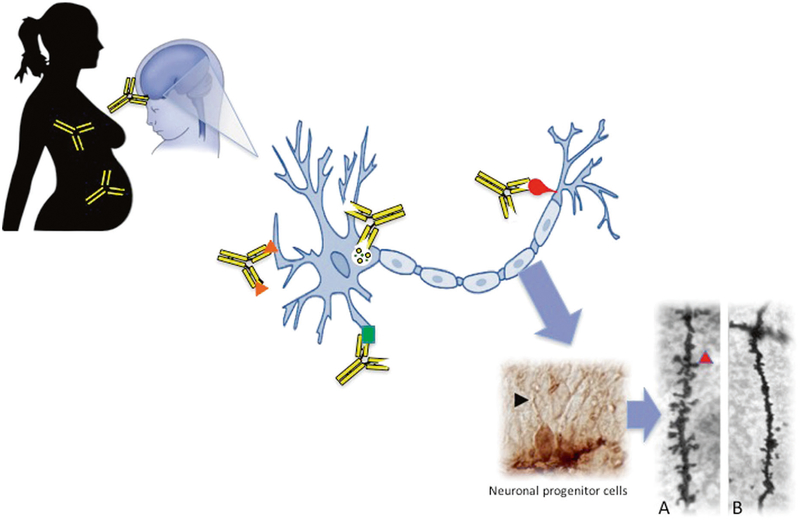
Fig. 2.
Proposed mechanism of maternal autoantibody related (MAR) ASD. Brain-reactive maternal autoantibodies cross the blood–brain barrier during gestation and bind to the various autoantigens on developing neurons (represented by orange triangles, green square, red bulb, and the intracellular antigen, LDH, by the yellow circles). We have shown that antibodies can bind intracellularly to neuronal progenitor cells in the developing fetal brain (black arrowhead) on embryonic day 14. Upon binding to their intracellular targets, these maternal autoantibodies elicit adverse effects to neurodevelopment such as a significant decrease in the number and density of dendritic spines on neurons in the developing cortex. This is illustrated in Panels a and b, as basal dendrites in frontal cortex layer V from an adult control mouse shows numerous dendritic spines (Panel a; red arrowhead), whereas offspring exposed on embryonic day 14 to human IgG with reactivity to LDH, CRMP1, and STIP1 lack mature spines (Panel b)

