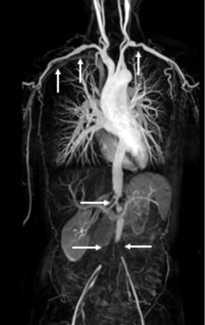
Figure 2.
Maximum intensity projection (MIP) reconstruction image from three-station magnetic resonance angiography (MRA) of a patient with Takayasu’s arteritis. The white arrows indicate areas of vascular pathology in the subclavian, axillary, abdominal, and iliofemoral arteries. Image from the National Institutes of Health Vasculitis Translational Research Program.

