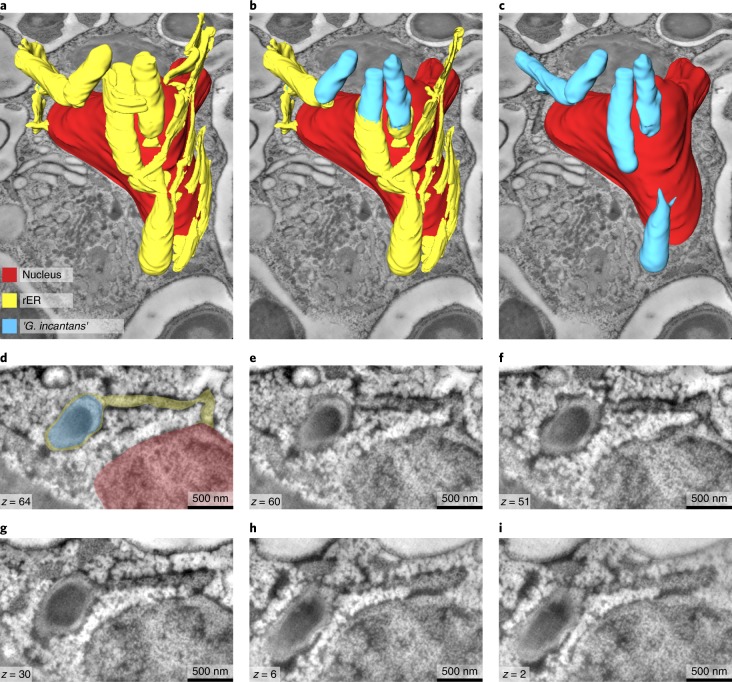
Fig. 3. ‘G. incantans’ lives in the rER of Trichoplax H2.
All panels are the results from a single experiment. a−c, 3D volume rendering of reconstructed ‘G. incantans’, the rER and the nucleus of a fibre cell, superimposed on a virtual slice of the 3D TEM tomography stack; the rER has been virtually removed to partially (b) and fully (c) show the symbionts within the rER lumen. No scale bar is shown as the scale varies with perspective. d−i, Selected tomography slices (on which the 3D reconstruction is based) at various depths (see z values indicated) show the connection between the nucleus, the rER and the bacteria. For ease of interpretation, d has been false-coloured using the colour key from a. For raw data, see Supplementary Fig. 8.

