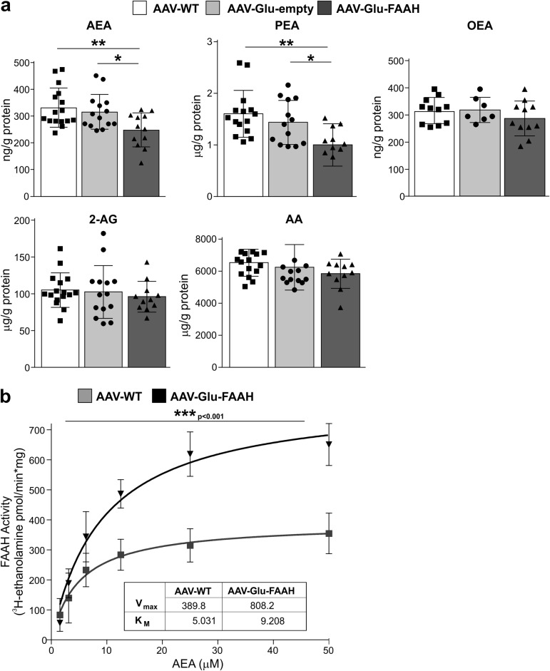
Fig. 2.
FAAH overexpression in hippocampal CA1-CA3 pyramidal neurons leads to decreased anandamide (AEA) and palmitoylethanolamide (PEA) levels. a AEA and PEA levels were significantly lower in AAV-Glu-FAAH samples (n = 11-12) as compared to control AAV-WT animals (n = 15-16) and AAV-empty injected mice (n = 12-14), whereas oleoylethanolamide (OEA), arachidonic acid (AA), and 2-arachidonoylglycerol (2-AG) levels were not altered. b FAAH activity is enhanced upon FAAH overexpression in hippocampus. Along increasing amounts of substrate [3H]-AEA (0–50 μM range), FAAH activity was increased in AAV-Glu-FAAH (n = 3) as compared to AAV-WT (n = 3) mice as indicated by the increase in the maximal amount of product [3H-ethanolamine], and the statistically significant increase in Vmax (inset). No significant change in Km constant was found. Data are expressed as amount of product [3H-ethanolamine] (pmol) generated over reaction time (min) and protein amount (mg). *p < 0.05, **p < 0.01, ***p < 0.001 one-way ANOVA with Tukey’s multiple comparison test. Vmax and Km values were obtained by using a Michaelis-Menten equation-based analysis. Values are expressed as means ± SEM. Statistical analysis details are reported in Supplementary Table S3.

