
Fig. 9.
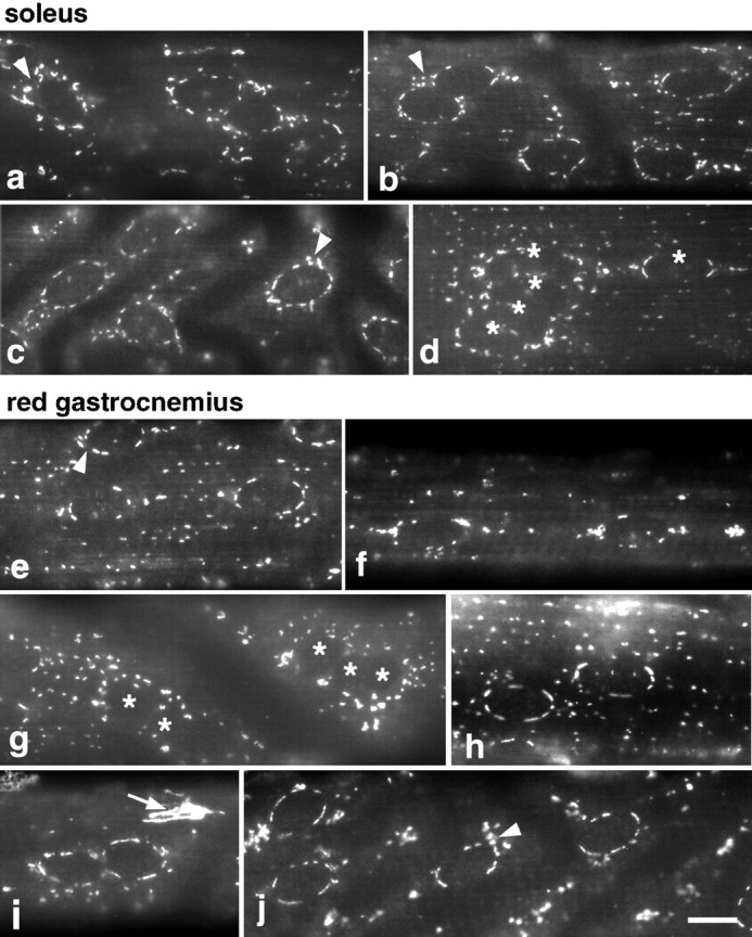
After denervation, the Golgi complex pattern is similar at the surface of type I and type II fibers. Five days after denervation, fibers from both denervated and contralateral control hindlegs of soleus and RG muscles were stained for MG160. The staining in the control fibers was similar to what is shown in Figure 1. In denervated soleus fibers (a–d), the perinuclear pattern is maintained. In RG fibers (e–j), all fibers have a perinuclear pattern that resembles that of the soleus (e, h–j). In some fibers, Golgi elements are still more concentrated at the nuclear poles (f). The perinuclear pattern is not as tight (arrowheads) as in the control fibers. dand g show aggregated nuclei, marked with anasterisk. Such aggregates are frequently encountered in the denervated but not in the control fibers (see Results). The bright Golgi complex of a nonmuscle cell is marked by a small arrow in i. Scale bar, 10 μm.