
Fig. 1.
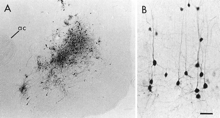
Light micrographs of a representative PRV injection in the NAc (A) and the resulting labeling of pyramidal neurons within the PFC (B). In A, the PRV injection is centered between the anterior commissure (ac) and the medial surface of the hemisphere. In B, many pyramidal neurons and their apical and basilar dendrites are extensively labeled after an injection of PRV into the NAc. Medial is toward the top, and dorsal is toward the left. Scale bar: A, 160 μm; B, 40 μm.