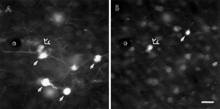
Fig. 2.
Fluorescence micrographs of the same coronal section through the PFC showing the distribution of neurons labeled for either PRV (A) or GABA (B) 48 hr after an injection of PRV into the NAc. Within this field, most neurons contain labeling for either PRV or GABA (small arrows), but not both markers. One neuron adjacent to a small blood vessel (asterisk) contains labeling for both markers (open arrow). Note the lighter fluorescence signal and limited dendritic labeling of the dual-labeled neuron compared with other PRV-labeled neurons within the same field. Scale bar, 20 μm.

