
Fig. 6.
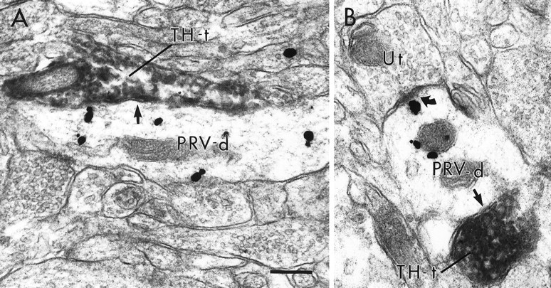
Electron micrographs of PRV-labeled dendrites contacted (straight arrows) by TH-immunoreactive terminals (TH-t) within the PFC. In A andB, PRV-labeled dendrites are contacted by TH-labeled terminals without forming distinct synaptic specializations. The PRV-labeled dendrite in B also receives an asymmetric synapse (curved arrow) from an unlabeled terminal (Ut). Scale bar, 0.25 μm.