
Fig. 5.
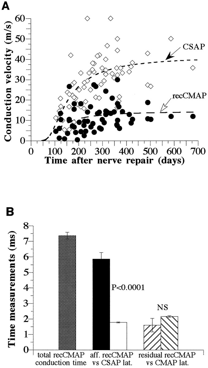
Conduction velocities (meters per second) and times (milliseconds) calculated from stimulating digital nerve at the tip or base of digit II. Data are from 11 median nerves (5 mm nerve lesion) and 131 physiological assessments. A, Conduction velocities of the CSAP and recCMAP along the same conduction path from the tip to the base of digit II as function of the time after nerve repair. The regression curves were calculated from the equation:a*exp(b/d2) using the least squares method (r = 0.4093 and 0.7387; p < 0.001). B, Estimates of conduction time, in milliseconds, of the recCMAP along the aberrant sensory (afferent) versus motor (efferent) pathways. Only time points >300 d after surgery were included (see A;n = 25 physiological assessments). The total conduction time for the recCMAP was calculated to be 7.4 ± 0.2 msec (mean ± SEM; hatched bar). The conduction time of the recCMAP along the sensory portion of the pathway (afferent;black bar), was calculated to be 5.9 ± 0.4 msec, which is significantly longer than the latency of the CSAP (in milliseconds) over the same pathway (white bar; 1.8 ± 0.03 msec; p < 0.0001, paired ttest). The residual conduction time of the recCMAP along the efferent motor pathway (calculated by subtraction of the afferent conduction time from the total conduction time) was not significantly different (paired t test) from the measured latency (in milliseconds) of the direct motor response evoked by stimulation at the wrist.