
Fig. 1.
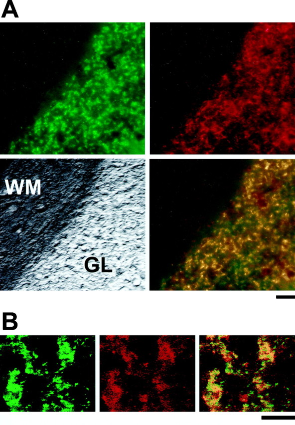
erbB4 and synaptophysin immunoreactivity colocalize at cerebellar glomeruli. A,Top, Parasagittal sections of adult cerebellum were stained with a monoclonal antibody to the synaptic vesicle protein synaptophysin (Oregon green secondary antibody; left) and polyclonal antibodies to erbB4 (0618) and visualized with a Cy3 secondary antibody (right). Bottom, The two top images are superimposed (right), and a bright-field image of the section is shown (left).B,Left, Middle, Confocal images were obtained at higher magnification of the granule cell layer from adult cerebellum stained as described above (synaptophysin,green; erbB4, red). Right, Colocalization of the two proteins is shown by the superimposition of the two images. GL, Granule cell layer; WM, white matter. Scale bars: A, 20 μm; B, 50 μm.