
Fig. 10.
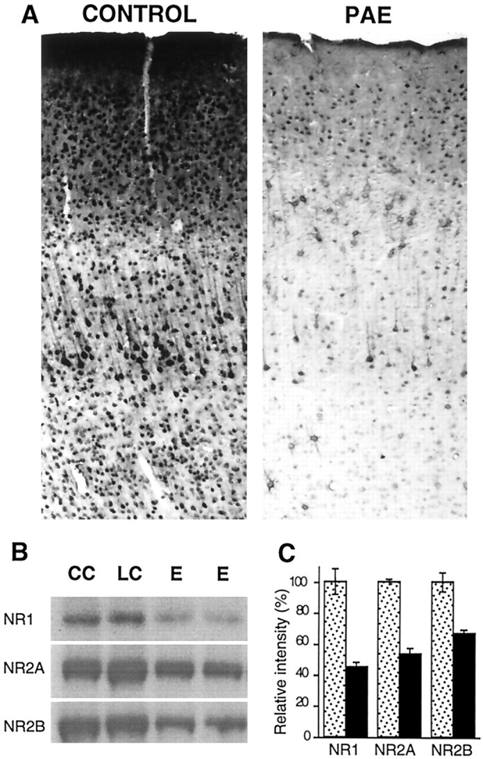
Analysis of NMDA receptor subunit levels after prenatal ethanol exposure. A, Immunohistochemical staining of the somatic sensory cortex from control and PAE rats with NR1 antibody AB59 showing a reduction in NMDAR1 protein in all layers. B, Western blot analyses of proteins from a membrane-enriched fraction of somatic sensory cortices of chow control (CC), liquid diet control (LC), and prenatal alcohol exposed (E) rats with antibodies to NR1(AB59), NR2A, and NR2B. Iodinated secondary antibodies were used to detect bands and to aid in quantification. C, The bands from the immunoblot were quantified using a phosphorimager, and the relative intensity refers to the values calculated by assigning arbitrary values of 100% to the controls.