
Fig. 15.
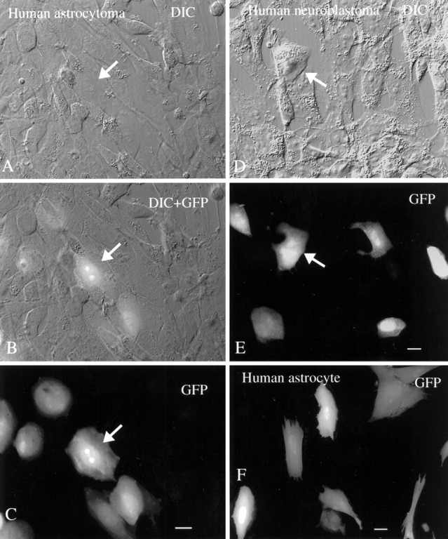
Human brain cells are infected with mouse CMV. A–C, DIC, DIC + GFP, and GFP images of the same microscope field containing glial cells from a human astrocytoma are shown. A subset of cells shows strong GFP-mediated fluorescence. Scale bar, 10 μm. D, E, The same field shows the DIC image (D) and the GFP image (E). A subset of human neuroblastoma cells, 2 d after plating with mCMV, is infected with virus. Scale bar, 10 μm. F, “Normal” glial cells with an astrocyte morphology were cultured from human brain and show GFP expression 30 hr after infection. Scale bar, 10 μm.