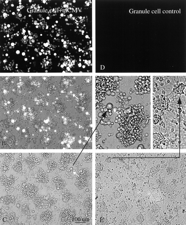
Fig. 8.

Cerebellar granule neurons. A, Four days after mCMV (MC.55) infection of a culture enriched with granule cells, many of the cells show bright GFP fluorescence. The same cell is indicated by an arrow in A–c'to facilitate recognition. B, Same field as inA but with partial fluorescence, partial phase contrast. Some cells seen in phase contrast are not fluorescent.C, Same field as in A but only with phase contrast. Scale bar, 100 μm. A higher magnification ofC is shown in c' (arrow). Neurites are not found in these infected neurons. D, Control granule cell culture not infected with mCMV shows no fluorescence. E, Phase-contrast photomicrograph of culture 2 d after infection. Neurites are commonly found spreading out from groups of neurons, shown in higher magnification ine'.
