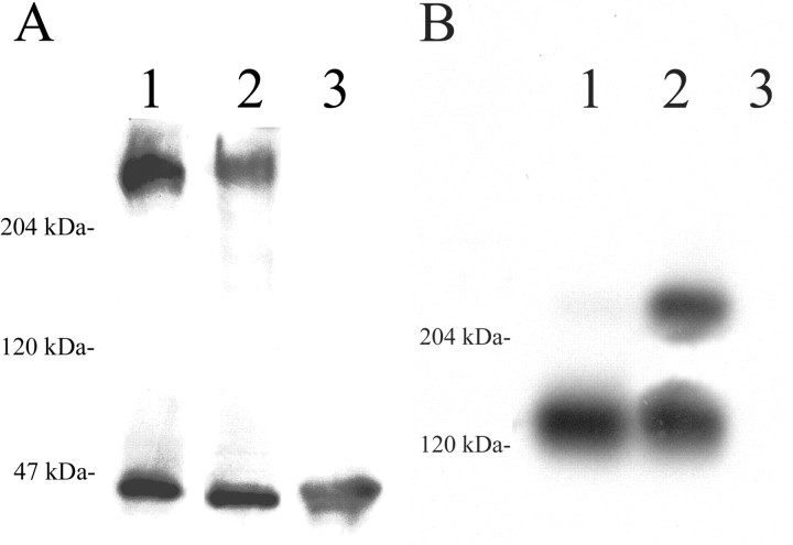
Fig. 6.
Immunoblot analysis of CS-PG expression in the glial scar. A, Levels of phosphacan protein in extracts prepared from gliotic tissue retrieved from filter implants (lane 2) are decreased to ∼67% of the levels in age-matched, uninjured cerebral cortex (lane 1), as detected with the 3F8 antibody. In this case, the phosphacan/MAPK values (in densitometric units) were 385,647/228,085 (ratio of 1.6881) and 228,141/202,899 (ratio of 1.1244) for uninjured cortex and filter implant, respectively. B, The 130 kDa proteolytic fragment of neurocan predominates in protein extracts from the cerebral cortex of uninjured age-matched control animals (lane 1), as detected using the 1F6 antibody to neurocan. Alternatively, in gliotic tissue retrieved from filter implants the full-length 245 kDa neonatal form of the neurocan protein is significantly upregulated (lane 2). In this case, the neurocan 245 kDa/neurocan 130 kDa values (see Materials and Methods) were 8454/378662 (ratio of 0.0223) and 284810/355734 (ratio of 0.8006) for uninjured cortex and filter implant, respectively. Neither phosphacan nor neurocan is detected in adult skeletal muscle (A and B,lane 3). ForA and B, 30 μg of protein were loaded in each lane.

