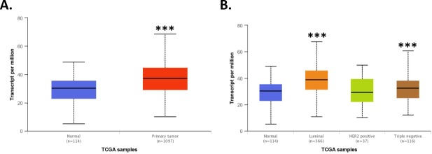
Fig 1. TCGA analysis of XRCC1.
A) TCGA analysis using the UALCAN web interface [23] revealed XRCC1 transcript expression to be significantly higher in breast tumor tissue over normal tissue (p = 1.6 x 10⁻¹², Primary tumor to Normal). B) XRCC1 was increased in Luminal (p < 1 x 10⁻¹², Luminal to Normal) and TNBC (p < 0.001, TNBC to Normal) tumor types compared to normal tissue using the same analysis interface. *** P < 0.001.

