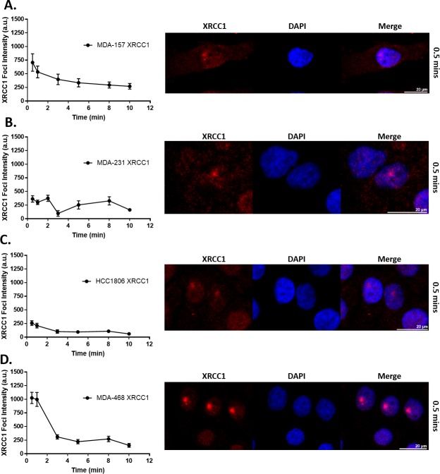
Fig 6. XRCC1 recruitment following 355 nm laser microirradiation in TNBC cell lines.
Microirradiation with a 355 nm laser occurred and cells were fixed at the indicated time points and processed for immunofluorescence of XRCC1. Quantification over time is shown on the left with a representative image from 0.5 min shown on the right for A) MDA-157, B) MDA-231, C) HCC1806, and D) MDA-468. A minimum of 18 cells were irradiated over three separate experiments and quantified as described in the materials and methods section and reported as XRCC1 Foci Intensity in arbitrary units (a.u.). Scale bar = 20 μm.

