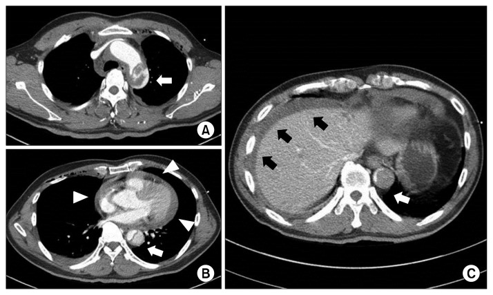
Fig. 1.
(A) Chest CT shows a traumatic aortic injury of the proximal descending thoracic aorta (white arrow). (B) Hemopericardium (white arrowheads) and dissection of the descending thoracic aorta are shown on CT. (C) Abdominal CT shows hemoperitoneum (black arrows) and dissection of the abdominal aorta (white arrow). CT, computed tomography.

