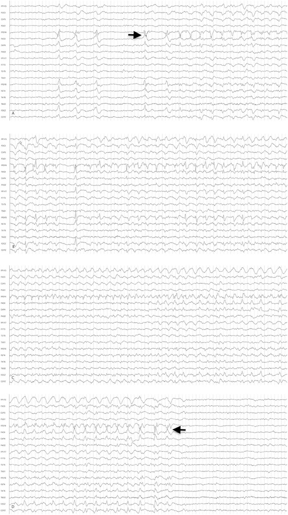
FIGURE 1:
Sample electrographic seizure. Electroencephalographic (EEG) example of a representative electrographic noncon-vulsive seizure from a 69-year-old male. An anterior-posterior bipolar montage is presented. (A) EEG shows sharp waves in the right hemisphere and seizure onset (arrow). (B) Seizure continuation. (C) Continuation of the seizure with maximum rhythmicity over the right anterior quadrant. (D) The termination (arrow) of the seizure. This seizure lasted about 50 seconds. Low-frequency filter = 1Hz; high-frequency filter = 70Hz; 60Hz filter is off.

