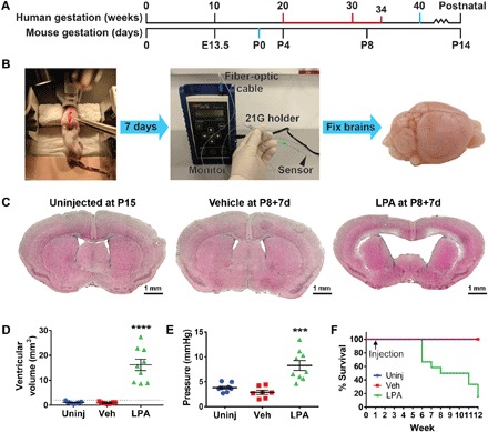
Fig. 1. Intracerebral LPA exposure induces hydrocephalus in neonatal mice.

(A) Approximate correlation of human and mouse brain development, highlighting full-term birth (40 weeks versus P0; blue) and ages at high risk for PHH (20 to 34 weeks versus P4 to P8+; red). (B) Experimental approach for this neonatal model of LPA-induced hydrocephalus. Mice received stereotactic intracranial injection at P8 (left; forceps point to approximate location of injection), followed by terminal ICP measurement (center) and brain harvest 7 days later (“P8+7d”; right). 21G, 21-gauge. (C) Intracerebral injection of LPA produces VM. Representative brain sections from uninjected (Uninj), vehicle-injected (Veh), and LPA-injected P8+7d mice quantified in (D). (D) Quantification of increased lateral ventricle volume (n = 10 per experimental group). The dotted line indicates 2 SDs above the vehicle mean. (E) Increased ICP in the brains of P8+7d mice injected with LPA (n = 9) compared to brains from uninjected (n = 8) or vehicle-injected (n = 7) mice. (D and E) Symbols indicate values from individual mice. ****P < 0.0001 and ***P < 0.0005 compared to vehicle controls. (F) Kaplan-Meier survival curve over a 12-week period for uninjected mice and mice injected with vehicle or LPA at P8 (n = 7 uninjected, n = 9 vehicle, and n = 11 LPA).
