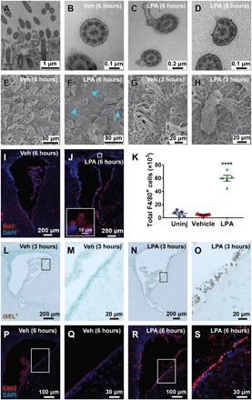
Fig. 3. Ependymal disruption precedes immune infiltration and cell death.

(A to D) TEM images of ependymal cilia cross sections. (A) Ciliary tuft with several distinct cilia adjacent to ependymal membrane. (B) Ciliary membrane and axoneme structure from a wild-type (WT) vehicle-injected mouse. (C) Disrupted ciliary membrane and (D) axoneme observed 6 hours after LPA exposure. (E to H) SEM images of the ventricular surface. (E) Intact ciliated ependymal surfaces were observed in vehicle-exposed brains. (F) LPA-exposed brain with ciliary loss and presence of cellular debris. The blue arrowheads point to remaining ciliary tufts 6 hours after LPA injection. (G) Vehicle-exposed cilia 3 hours after injection. (H) Possible phagocyte observed 3 hours after LPA exposure attached to shriveled and disorganized ependymal cilia. (I and J) Immunofluorescence images of Iba1+ innate immune cells in the lateral ventricle 6 hours following (I) vehicle or (J) LPA injection (×10 with ×63 inset). (K) Total number of F4/80+ CD11b+ Ly6G− macrophages and microglia isolated from brains of uninjected, vehicle-injected, or LPA-injected mice 24 hours after injection, as determined by flow cytometry. ****P < 0.0001 compared to vehicle controls. n = 5 individual brains combined from two independent experiments. (L to O) In situ end-labeling plus (ISEL+) assay indicating DNA damage 3 hours after injection (brown, fragmented DNA; light green, nuclei). (L and M) Brains exposed to vehicle were unaffected, whereas (N and O) LPA exposure caused selective ependymal DNA damage, with (L and N) ×10 magnification and (M and O) boxed areas enlarged to ×63. (P to S) Cleaved caspase 3 (Cas3) (red, with blue nuclear counterstain) immunolabeling in (P and Q) vehicle-injected and in (R and S) LPA-injected brains, which show selective ependymal apoptosis 6 hours following LPA exposure. (P and R) Images at x20 magnification with (Q and S) boxed areas enlarged to 63×.
