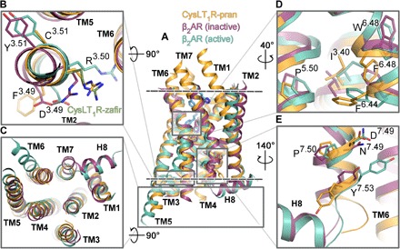
Fig. 2. Functional motifs of CysLT1R show unusual inactive-state features.

(A) Superposition of CysLT1R-pran (orange) with β2AR in inactive (PDB ID 2RH1; violet) and active (PDB ID 3SN6; teal) conformations. The membrane boundaries are shown as dashed lines. Loops are removed for clarity. (B to E) Zoom in on functional elements: DRY motif (B), intracellular region (C), P-I-F motif (D), and NPxxY motif (E). A different conformation of R1213.50 in CysLT1R-zafir (chain A) is shown as green sticks in (B).
