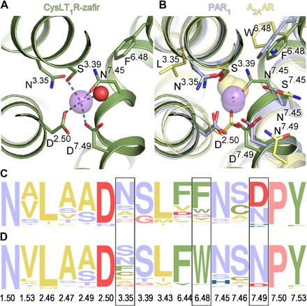
Fig. 3. Sodium-binding pocket in CysLT1R.

(A) Details of Na+ (purple sphere) coordination. Water molecule is shown as a red sphere. (B) Comparison of all high-resolution GPCR structures with resolved Na+. Sodium ions are shown as purple spheres for the δ-branch receptors: CysLT1R-zafir (green), PAR1 (light purple; PDB ID 3VW7), PAR2 (PDB ID 5NDD), and as yellow spheres for receptors from the α-branch: A2AAR (yellow; PDB ID 4EIY) and β1AR (turkey; PDB ID 4BVN), and the γ-branch: DOR (PDB ID 4N6H). (C and D) Frequency analysis of amino acid occurrence in the sodium pocket of the δ-branch class A GPCRs (C) and other class A receptors excluding the δ-branch (D). Yellow color marks amino acids with hydrophobic side chains; green, aromatic; red, negatively charged; blue, positively charged; purple, polar uncharged; pink, Gly and Pro. Frames indicate positions with the largest differences. The frequency analysis was performed using the weblogo.berkeley.edu server.
