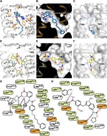
Fig. 4. Orthosteric ligand-binding pocket in CysLT1R.

(A and D) Details of ligand-receptor interactions for pranlukast (A) and zafirlukast (D). (B and E) Pocket shapes for pranlukast (B) and zafirlukast (E). (C and F) Pocket entrance for pranlukast, closed “gate” (C), and zafirlukast, open “gate” (F). (G and H) 2D representations of receptor-ligand interactions for pranlukast (G) and zafirlukast (H). Water molecules are shown as red spheres in (A). Residues engaged in the same type of interactions with both zafirlukast and pranlukast are colored in light green, and those engaged in different types are colored in orange (G and H). The membrane boundary is shown as a dashed line in (B, C, E, and F).
