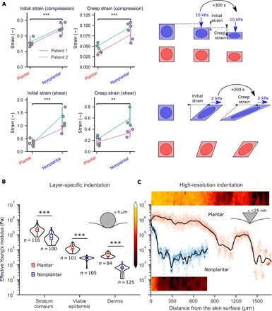
Fig. 3. Plantar skin resists deformation.

(A) Uniaxial compression and simple shear tests on ex vivo skin. Deformation was measured as the initial strain after compression of 10 kPa (top) and shear of 2 kPa (bottom). Tests are from two patients, with three samples from each anatomical location. Loads were maintained for 300 s, and final deformation was measured as creep strain. (B) AFM indentation experiments using a spherical (4 μm) tip on cryostat sections of skin. (C) High-resolution force mapping using a sharp AFM tip shows that the change in Young’s modulus with depth is more gradual in plantar skin. Two-dimensional (2D) stiffness maps (50 μm wide) are shown alongside depth-specific data. Black lines represent LOESS regression fits of the data, while color intensity represents the spread of elastic moduli at each depth. ***P < 0.001, **P < 0.01, two-sided Student’s t test.
