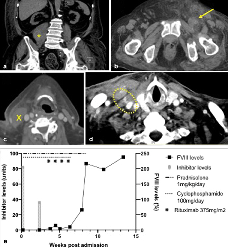
Fig. 1.

Simultaneous presentation of bleeding and thrombosis. ( a, b ) Noncontrast CT scan of abdomen and pelvis demonstrating ( a ) right psoas swelling, depicted with star , and ( b ) obliteration of the left femoral vein ( arrow ); ( c, d ) CT neck with contrast demonstrating ( c ) a hematoma overlying the right sternocleidomastoid ( cross ) and ( d ) contrast extravasation from the right IJV puncture site; ( e ) Serum FVIII and inhibitor levels with respect to immunosuppression therapy. CT, computed tomography; IJV, internal jugular vein.
