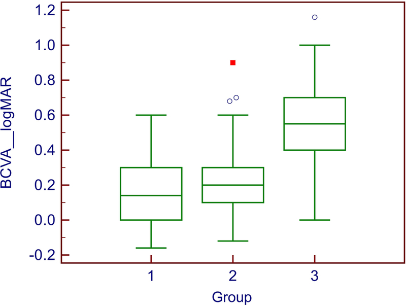
Figure 5: Distribution of BCVA values by location of the EZ break.

The differences in BCVA between groups 1 (no EZ break, n=24) and 2 (EZ break present but not involving the foveal center, n=93) were not statistically significant. In group 3 (n=43), two eyes had full vision. One explanation of good visual acuity despite the lesion reaching the foveal center in these outliers may be that fixation and the point of best acuity may not correspond exactly with the anatomical center of the fovea in all eyes.(Zeffren et al. 1990; Wilk et al. 2017; Putnam et al. 2005) Another possibility is that the absence of the EZ line may not indicate absence of structures that give rise to the line, but indicate distortion of alignment of these structures.
