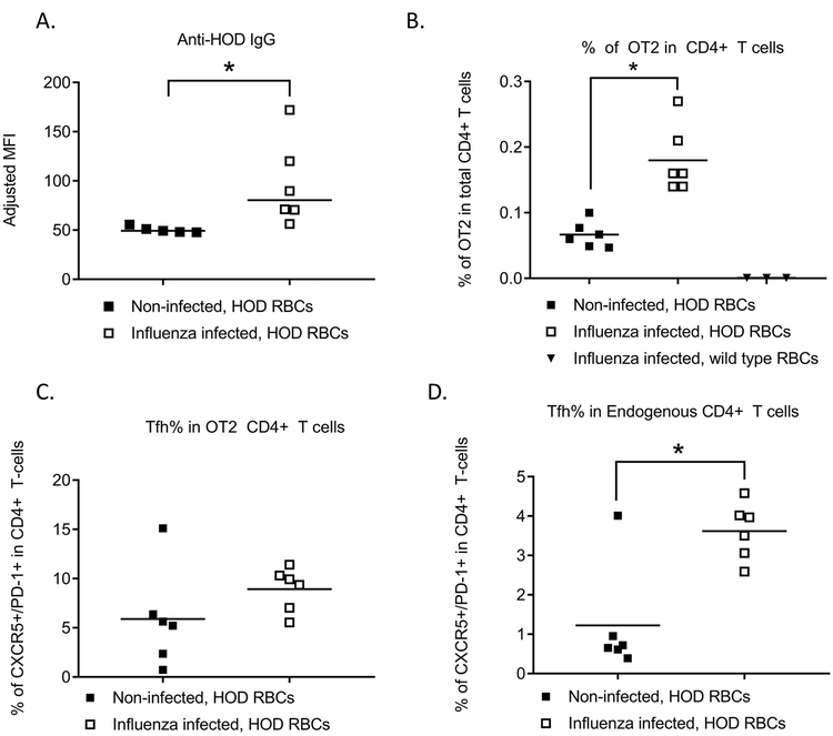
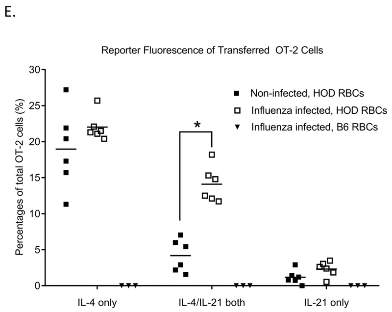
Figure 2. Influenza infection promotes antigen-specific Tfh generation and dual IL4/IL21 production.
104 OT-2 CD4+ T-cells were adoptively transferred into naïve wild type C57BL/6 mice or mice infected with 10 PFU of PR8 influenza virus, followed by a HOD RBC transfusion. (A) Anti-HOD IgG alloantibody responses as measured by flow cytometry. (B) Percentage of OT-2 cells in total CD4+ T-cells, (C) percentage of endogenous CD4+ T-cells that were CXCR5+/PD-1+, and (D) percentage of OT-2 CD4+ T-cells that were CXCR5+/PD-1+ were evaluated by flow cytometry 6 days post-RBC transfusion. For (E), 104 cells CD4+ T-cells from C57BL/6 (B6) IL21Kat/+IL4GFP/+ double reporter mice crossed with OT-2 mice were adoptively transferred into naïve wild type C57BL/6 mice or mice infected with 10 PFU of PR8 influenza virus, followed by a HOD RBC transfusion. Fluorescence of IL21 and IL4 producing cells was evaluated 6 days post-transfusion, after gating on ova-specific CD4+ T-cells. For B-E, pre-gating included inclusion of TCRβ+ cells, exclusion of B220+ cells, and inclusion of CD4+, CD44+ cells. OT-2 cells were identified by CD45.1 or CD90.1 positivity. Data shown are representative of 3 independent experiments, *p<0.05.


