
Figure 3.
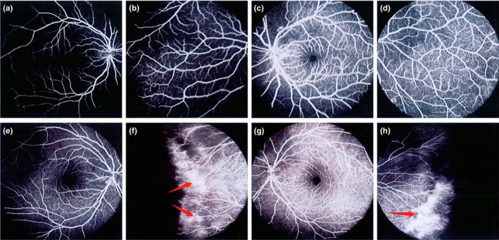
Subject III‐1: (a, b) represent right eye; (c, d) represent left eye. Fluorescein fundus angiography showed normal appearance of the posterior pole and peripheral retinal vessels of the right eye and left eye of the proband's father (III‐1), respectively; Subject III III‐2: (e, f) represent right eye; (g, h) represent left eye. (e, g) Respectively showed that the mother (III‐2) of the proband had normal fundus of the posterior pole of the right eye and the left eye. (f, h) Respectively showed that the mother of the proband had abnormal manifestations of peripheral retinal vessels in the right eye and left eye. Fluorescent fundus angiography showed no perfusion area in the peripheral retina, and fluorescence leakage appeared in the peripheral blood vessels of the retina