Figure 2.

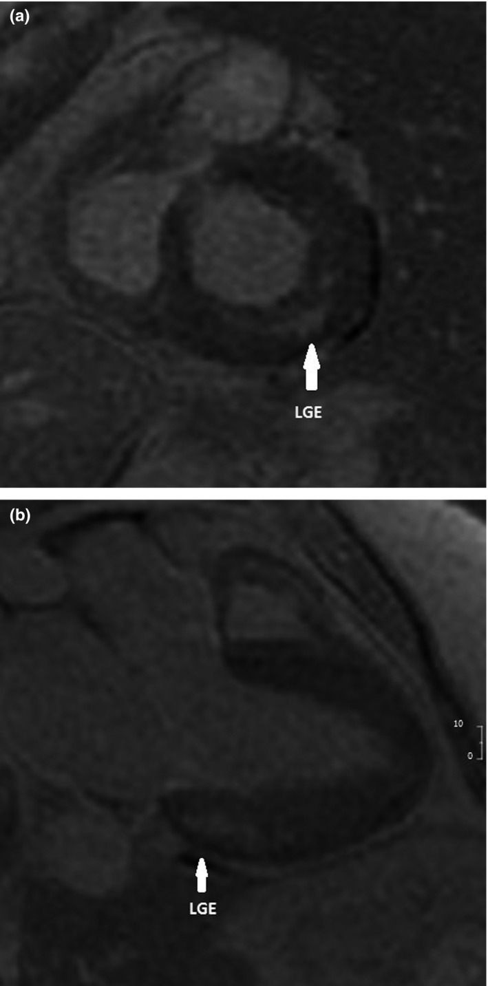
Short‐axis (a) and long‐axis (b) delayed enhanced images showing mid‐wall enhancement (white arrows) in the inferolateral wall of the left ventricle

Short‐axis (a) and long‐axis (b) delayed enhanced images showing mid‐wall enhancement (white arrows) in the inferolateral wall of the left ventricle