Fig. 8.
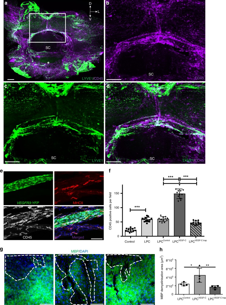
Interactions of spinal LVs with immune cells. a–d Double labeling of cleared cervical vertebral column segments with antibodies against LYVE1 (green) and CD45 (purple), spatial orientation (D: dorsal, L: lateral, V: ventral). b–d Magnified images of white box in (a). Merged images (a), (d) show CD45+ leukocytes located along LYVE1+ vLVs. White asterisk: vertebral ventral body; SC: Spinal cord. e Cryosection of a cervical vertebra from a Vegfr3:YFP mouse labeled with antibodies against MHCII (red) and CD45 (white). CD45+ leukocytes including MHCII+ antigen-presenting cells are located close to and inside a YFP+ vLV (green) in the ligament flavum. f Quantification of CD45+ cells in vertebral column whole-mount preparations (see stippled area in Fig. 7i). g Cryosections of the lumbar spinal cord from LPC-injured mice previously injected with AAV-VEGFR34–7-Ig (LPCcontrol), AAV-mVEGF-C (LPCVEGF-C) or AAV-mVEGFR-31–3-Ig (LPCVEGF-C trap) in the lumbo-sacral region. Images representative of the ipsilateral side showing MBP+ myelin (green) and demyelinated area (dashed lines) with Hoechst+ nuclear staining (blue) in (g). h Histograms showing quantification of MBP-negative demyelinated area (dotted line in (g)) at the lesion site. Demyelinated area is increased in LPCVEGF-C mice compared to LPCcontrol mice. n = 4 biologically independent mice/independent experiment, and data represent mean+/−SD (error bar); one-way ANOVA with Tukey’s multiple-comparisons test; *p < 0.05, ***p < 0.001. Source data are provided as a Source Data file. Scale bars: 300 µm (a–d); 50 µm (e); 100 µm (g)

