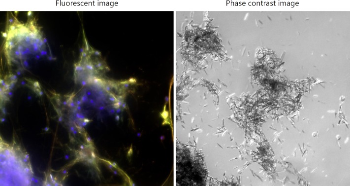
Fig. 3.
Neutrophil death with DNA release and decondensation triggered by monosodium urate crystals. Healthy control neutrophils incubated with monosodium urate crystals (0.2 mg/mL) for 4 h. Maximum-intensity projection of a z-stack. The fluorescent image is a merge of blue (DAPI), green (neutrophil elastase) and red (myeloperoxidase). DNA colocalised with neutrophil granule proteins aggregates the crystals.

