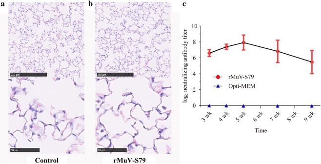
Fig. 3.
Cotton rats were grouped infected with 106 PFU of rMuV-S79. Five cotton rats were inoculated Opti-MEM as control group. Histological examination was conducted by HE staining of lung tissues from vaccination group (a) and control group (b) at 4th day post-inoculation. Sera were obtained at 3–5, 7, and 9 weeks after immunization to detect neutralization anti-MuV antibodies. The antibodies titers at each time point quantified by 50% plaque reduction assay are calculated and shown as mean NT titers (c)

