. 2019 Sep 2;18:750–763. doi: 10.17179/excli2019-1459

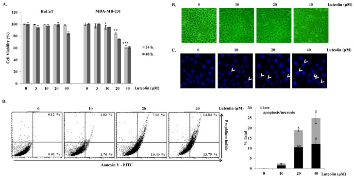
Figure 3. Cytotoxic and apoptotic effects of luteolin in MDA-MB-231 breast cancer cells. (A) MDA-MB-231 breast cancer cells and HaCaT normal keratinocytes were treated with luteolin for 24 or 48 h. Cell viability was determined by MTS assay. (B) Morphological changes in MDA-MB-231 cells after treatment with various concentrations (0, 10, 20, and 40 μM) of luteolin for 24 h were observed under a phase-contrast microscope (100×). (C) Apoptotic nuclei after treatment with luteolin were observed under a fluorescence microscope after Hoechst staining (100×). (D) Flow-cytometric analysis after staining with Annexin V-fluorescein isothiocyanate (FITC) and propidium iodide (PI). Percentages of significant events in early apoptosis (bottom right quadrants) and late apoptosis (top right quadrants). Bar graph represents annexin V+/PI- (early apoptotic) and annexin V+/PI+ (late apoptotic) cells (n = 3). Statistically significant differences between luteolin-treated versus non-treated cells were analyzed by two-tailed Student's t-test. *p < 0.05, **p < 0.005, ***p < 0.001 (n = 3).

Figure 3