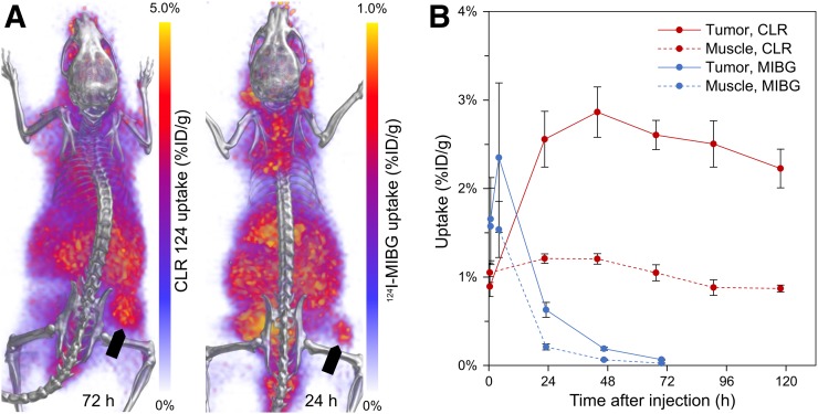
FIGURE 5.
Whole-body PET/CT imaging and quantitative analysis of tumor-specific uptake of CLR 124 and 124I-MIBG in murine xenograft models of NB1691-hNET. (A) Representative murine xenograft models of NB1691-hNET administered CLR 124 or 124I-MIBG are presented as 3-dimensional renderings of PET (purple and yellow) fused with CT (grayscale) images acquired during peak tumor-specific uptake at 72 and 24 h after injection, respectively. Location of flank tumor is indicated by arrows. (B) PET/CT ROI analysis of tumor and contralateral muscle uptake is shown with mean values (n = 4) and error bars for SE.

