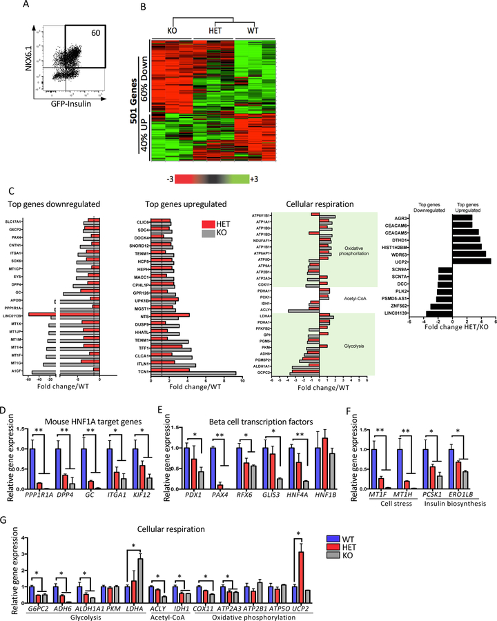
Figure 5: Disruption of HNF1A leads to an abnormal expression of genes related with beta cell function, development and diabetes.
Mel1 HNF1A WT, HET and KO ESCs were differentiated into pancreatic endocrine cells and analyzed as described below.
(A–B) HNF1A allelic series genome profiling using purified INS-GFP+ NKX6.1+ cells at end stage of differentiation (n=3 per sample). (A) Flow cytometry plot for sorting double positive INS-GFP+ NKX6.1+ cells. (B) 501 differential expressed genes presented in a heat map plot obtained using a microarray analysis from samples sorted in A.
(C) Differential expressed genes shown in the following categories: top downregulated genes, top upregulated genes, cellular respiration and genes differentially expressed in HET compared to KO.
(D–G) qRT-PCR gene expression validation of a subset of gene targets in B (n=3 per sample). (D) Mouse HNF1A target genes. (E) Pancreas transcription factors. (F) cell stress and insulin biosynthesis. (G) Cellular respiration.
For all statistical analyses: * P<0.05, **P<0.01. See also Figure S5.

