Figure 3.
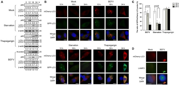
BEFV promotes autophagosome formation. A MDBK cells were respectively infected with BEFV at a MOI of 1, starved (cells supplemented with no FBS), or treated with thapsigarigin (10 µM). The cell lysates were collected at the indicated time points and subjected to immunoblotting with the respective antibodies. Signals were quantified using Image J software. The levels of the indicated proteins at 0 h were considered onefold. The protein levels were normalised to those for β-actin. The activation and inactivation folds indicated below each lane were normalized against values for the 0 h. The predicated size of each protein was labeled at the right-hand side in kDa. B LC3 puncta were observed under the fluorescence microscope. Cell nuclei were stained by DAPI. Scale bars, 25 µm. Normalization of the GFP/mCherry-LC3 puncta (%) were presented in C. D MDBK cells were transfected with mCherry-LC3 plasmid for 24 h and then infected with BEFV at MOI of 1 for 18 h. The cells were then fixed and processed for immunofluorescence staining of mCherry-LC3 and LAMP2. The colocalization of LAMP2 and LC3 was observed under a fluorescence microscope. Scale bar, 25 µm.

