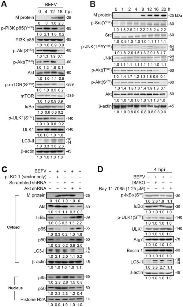
Figure 4.

BEFV induces autophagy through upregulation of the PI3K/Akt/NF-κB pathway in the early to middle stages. A, B MDBK cells were infected with BEFV at a MOI of 1 and the cell lysates were collected individually at the indicated time points for immunoblotting with the indicated antibodies. C The levels of Akt, IkBα, p65, p50, and LC3-II were examined in BEFV-infected cells transfected with an Akt shRNA. D MDBK cells were pretreated with NF-κB inhibitor Bay 11-7085 and the cell lysates were collected individually at 4 hpi for immunoblotting with the respective antibodies. Signals were quantified using Image J software. The levels of the indicated proteins in the mock controls (0 h or un-treatment) were considered onefold. The protein levels were normalised to those for β-actin. The activation and inactivation folds indicated below each lane were normalized against values for the mock control. The predicted size of each protein was labeled at the right-hand side in kDa.
